產業類別名稱
 The LVEM5 multimodal imaging capabilities makes it a comprehensive imaging tool. The LVEM5 is truly a 3-in-1 electron microscope. Not only is it a Transmission Electron Microscope (TEM), but it can be configured with up to two different scanning modes for use as a Scanning Election Microscope (SEM) and a Scanning Transmission Electron Microscope (STEM). With the LVEM5 you can switch between imaging modes without moving your sample. This way you can capture both surface and transmission images from the same area of interest. With only one tool you can significantly improve the understanding of various nanoparticles.
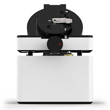
The LVEM5 is the most cost-effective multi-modal electron microscope available in a benchtop configuration. The LVEM25 is built upon the same platform as the LVEM5, but operates at a slightly higher accelerating voltage allowing for an improvement in final image resolution. Both LVEM microscopes miniature size means that they can be installed in your workspace, right where you need it. Don't let the small size of the LVEM microscopes mislead you. The LVEM5 is capable of resolving objects as small as 1.2 nanometers in transmission modes, while the LVEM25 can resolve features down to 1.0nm. Additionally, the LVEM systems are capable of producing higher contrast images than conventional transmission electron microscopes without the need for stain.
In no way are you sacrificing imaging quality or obtainable resolution with a benchtop configuration. The LVEM microscopes easily produce high quality images suitable for presentations or publications. They are also so remarkably simple that anyone can use one. Whether it is for clusters, precipitates, nanowires, nanorods, or quantum dots, particle size can be estimated immediately with a LVEM on a live image. It can then be measured systematically on collected images and the data exported to Excel or statistics package of choice.